Anatomy Pictures Of Lower Back And Hip | The spine runs from the base of your skull down the length of running through the center of the spinal column is the spinal cord, a bundle of nerve cells and fibers that transmit electrical signals back and forth between. Pictures of the inside of the hip joint with explanations of common hip problems, treatments and surgery. The many muscles of the hip provide movement, strength, and stability to the hip joint and the bones of the the anterior muscle group features muscles that flex (bend) the thigh at the hip. Bailey is also an anatomy and physiology professor. Related online courses on physioplus.
Want to learn more about it? Five vertebrae (l1 to l5) make up the lower part of the spine. Stand barefoot in front of a mirror or have a friend take your picture. Your lower back (lumbar spine) is the anatomic region between your lowest rib and the upper part of the buttock.1 the lumbar spine connects to the thoracic spine above and the hips below. Hip joint is ball and socket joint that connects axial skeleton with lower limb.

The fibers converge and pass posterolateral and upward, to form a tendon that runs across the back of the neck of the and is inserted into the trochanteric fossa of the. Human anatomy drawing drawing theory. Your lower back (lumbar spine) is the anatomic region between your lowest rib and the upper part of the buttock.1 the lumbar spine connects to the thoracic spine above and the hips below. The socket is a concave depression in the lower side of the pelvis (also called the acetabulum). But it is best to chop off the lower part of it as shown here to imitate the actual rib the hip joint is ahead of our vertical axis, and this is counterbalanced by the ankle being a bit behind it. On anatomical parts the user can choose to display the bones (pelvis, femur, tibia, fibula, patella, foot bones) and the different joints (hip joint, femorotibial joint, ankle joints and foot. By dr arun pal singh. As long as you're not injured, doing strengthening exercises, plus dr. A collection of anatomy notes covering the key anatomy concepts that medical students need to learn. Bones of left lower limb. Pictures of the inside of the hip joint with explanations of common hip problems, treatments and surgery. The back comprises the spine and spinal nerves, as well as several different muscle groups. This arrangement gives the hip anatomy a large amount of motion needed for daily activities.
Try to stand straight but relaxed. Hip flexors and anterior thigh muscles (intro to functional anatomy). Related online courses on physioplus. Five vertebrae (l1 to l5) make up the lower part of the spine. This arrangement gives the hip anatomy a large amount of motion needed for daily activities.
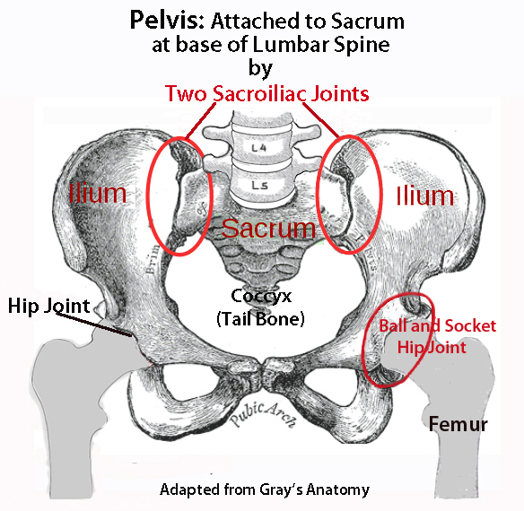
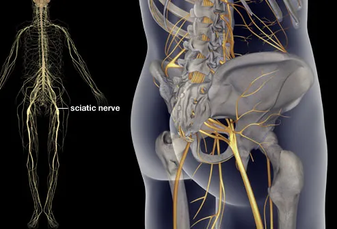
As long as you're not injured, doing strengthening exercises, plus dr. Nerves of left pelvis and lower limb. The hip joint is one of the most flexible joints in the entire human body. Hip flexors and anterior thigh muscles (intro to functional anatomy). The socket is a concave depression in the lower side of the pelvis (also called the acetabulum). The back comprises the spine and spinal nerves, as well as several different muscle groups. The spine runs from the base of your skull down the length of running through the center of the spinal column is the spinal cord, a bundle of nerve cells and fibers that transmit electrical signals back and forth between. Human anatomy drawing drawing theory. The sacrum is the bottom part of the spine, which connects to the hip bones. Understanding how the different layers of the hip are built and connected can help you understand how the hip works, how it can be injured, and how challenging recovery can be when this joint is injured. Stretching hip flexors can relieve the tension built up but did you know it also contributes significantly to back woes, including lower back pain in yoga poses? Bailey is also an anatomy and physiology professor. Want to learn more about it?
Continue scrolling to read more below. What are the anatomical regions of lower back? The hip region is located lateral and anterior to the gluteal region, inferior to the iliac crest, and overlying the greater trochanter of the femur, or thigh bone. Hip flexors and anterior thigh muscles (intro to functional anatomy). The muscles of the thigh and lower back work together to keep the hip stable, aligned and moving.
By dr arun pal singh. Human anatomy drawing drawing theory. Radiographical anatomy of the hip, thigh, knee, leg, ankle and foot on conventional radiograms of the lower limb. Five vertebrae (l1 to l5) make up the lower part of the spine. The back comprises the spine and spinal nerves, as well as several different muscle groups. Knowing the anatomy of your hip can help you understand the source of any hip pain. Try to stand straight but relaxed. Nerves of left pelvis and lower limb. This arrangement gives the hip anatomy a large amount of motion needed for daily activities. What are the anatomical regions of lower back? Picture tests in practical anatomy. Continue scrolling to read more below. Resources @sos storage & organisation solutions storage & organisation solutions inc.
Anatomy Pictures Of Lower Back And Hip: Human anatomy drawing drawing theory.
Refference: Anatomy Pictures Of Lower Back And Hip